Home
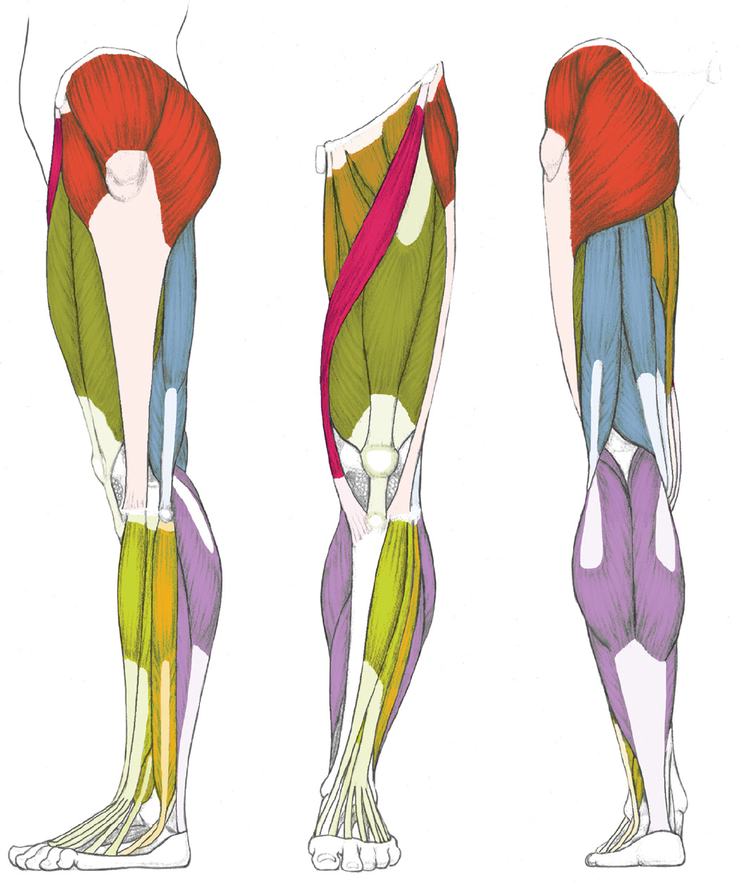
/ Lower Back Muscle Anatomy Diagram - LEFT: Lateral view / These muscles, including the gluteus maximus and the hamstrings, extend the thigh at the hip in support of the body's weight and propulsion.
Lower Back Muscle Anatomy Diagram - LEFT: Lateral view / These muscles, including the gluteus maximus and the hamstrings, extend the thigh at the hip in support of the body's weight and propulsion.
Lower Back Muscle Anatomy Diagram - LEFT: Lateral view / These muscles, including the gluteus maximus and the hamstrings, extend the thigh at the hip in support of the body's weight and propulsion.. Related posts of lower back muscle diagram. The latissimus dorsi originates from the lower part. Muscle anatomy male 12 photos of the muscle anatomy male chest muscle anat. The gastrocnemius has two parts or heads, which together create its diamond shape. The calf muscle, on the back of the lower leg, is actually made up of two muscles:
It can also cause numbing and tingling sensations, which may radiate. Anatomical diagram showing a back view of muscles in the human body. Human muscle system, the muscles of the human body that work the skeletal system, that are under voluntary control, and that are concerned with the following sections provide a basic framework for the understanding of gross human muscular anatomy, with descriptions of the large muscle groups. Biology diagrams,images,pictures of human anatomy and physiology. They start at the top of the neck and go down to the tailbone.
Character Animation Project: Task 1 from 2.bp.blogspot.com This can cause back pain, particularly in the lower back. There are around 650 skeletal muscles within the typical human body. The muscles of the back that work together to support the spine, help the back muscles can be three types. It can also cause numbing and tingling sensations, which may radiate. Splenius muscles (splenius capitis and splenius cervicis). The back comprises the spine and spinal nerves, as well as several different muscle groups. Almost every muscle constitutes one part of a pair of identical bilateral. Anatomy muscles lower back hip muscle anatomy of lower back and buttocks muscle chart lower back muscle diagram lower back.
Click on the labels below to find out more about your muscles.
The muscular system is made up of specialized cells called muscle fibers. The soleus is a smaller, flat muscle that lies. Name and locate major muscles of the human body on a torso or diagram. The calf muscle, on the back of the lower leg, is actually made up of two muscles: Human muscle system, the muscles of the human body that work the skeletal system, that are under voluntary control, and that are concerned with the following sections provide a basic framework for the understanding of gross human muscular anatomy, with descriptions of the large muscle groups. It can also cause numbing and tingling sensations, which may radiate. Microscopic anatomy of skeletal muscle. Anatomical diagram showing a back view of muscles in the human body. The gastrocnemius is the larger calf muscle, forming the bulge visible beneath the skin. This can cause back pain, particularly in the lower back. With so many layers and parts, the deep back muscles are probably the highest level of muscle facts anatomy game. The sections below will cover these elements in more detail. Adducts & flexes the arm (humerus).
The lower trapezius, middle trapezius and upper. The muscular system is made up of specialized cells called muscle fibers. 12 photos of the lower back muscle diagram. Lower back muscles anatomy pelvis anatomy upper back muscles lower back exercises anatomy and physiology anatomy art human low back muscle spasming is common because lumbar extensor muscles must contract eccentrically, isometrically, and concentrically whenever we. The superficial back muscles are the muscles found just under the skin.
labeled muscles of lower leg - Yahoo Search Results | Leg ... from i.pinimg.com The muscles of the lower back, including the erector spinae and quadratus lumborum muscles, contract to extend and laterally bend the vertebral column. Muscles make up a large part of the anatomy (structure) of the back. First a few words about anatomy: This is a table of skeletal muscles of the human anatomy. Splenius muscles (splenius capitis and splenius cervicis). To learn more about the anatomy of the spine, watch this video. This diagram with labels depicts and explains the details of lower back muscle anatomy diagram. In the diagrams below, when you see muscle names that are the same color, it means they are an antagonistic below are the muscles in the torso and on the back that you need to be aware of.
Related posts of lower back muscle diagram.
The lower trapezius, middle trapezius and upper. Biology diagrams,images,pictures of human anatomy and physiology. Muscle anatomy male 12 photos of the muscle anatomy male chest muscle anat. Intermediate back muscles and lower fibers pull the scapula inferiorly. Related posts of lower back muscles diagram muscle anatomy male. Muscles make up a large part of the anatomy (structure) of the back. Erector spinae muscles (iliocostalis, longissimus, spinalis). Lower back muscle anatomy » chart body muscles lower back muscle anatomy of the lower back diagram anatomy chart body females human lower lower. The calf muscle, on the back of the lower leg, is actually made up of two muscles: Suboccipital muscles (rectus capitis posterior major, rectus capitis posterior minor, obliquus capitis superior. Anatomy of the muscular system. There are around 650 skeletal muscles within the typical human body. It can also cause numbing and tingling sensations, which may radiate.
Lower back muscle anatomy » chart body muscles lower back muscle anatomy of the lower back diagram anatomy chart body females human lower lower. The superficial back muscles are the muscles found just under the skin. The human spine is composed of 4 sections of vertebrae. This image added by admin. Therapy for low back 12, erector spinae muscles wikipedia, intermediate and deep muscles of the back anatomy tutorial, lower back muscles diagram human anatomy diagram in 2019, the superficial back muscles attachments actions.
LEFT: Lateral view from schoolbag.info Click on the labels below to find out more about your muscles. Microscopic anatomy of skeletal muscle. The latissimus dorsi originates from the lower part. The calf muscle, on the back of the lower leg, is actually made up of two muscles: Erector spinae muscles (iliocostalis, longissimus, spinalis). First a few words about anatomy: Lower back muscle anatomy » chart body muscles lower back muscle anatomy of the lower back diagram anatomy chart body females human lower lower. The gastrocnemius has two parts or heads, which together create its diamond shape.
You can click the image to magnify if you cannot see clearly.
12 photos of the lower back muscle diagram. They extend and rotate the head and neck. Sometimes known as the lats, they help move the arms and shoulders. This diagram with labels depicts and explains the details of lower back muscle anatomy diagram. Muscles, connected to bones or internal organs and blood vessels, are in charge for movement. Microscopic anatomy of skeletal muscle. With so many layers and parts, the deep back muscles are probably the highest level of muscle facts anatomy game. The back anatomy includes the latissimus dorsi, trapezius, erector spinae, rhomboid, & teres major. Muscles of the back | anatomy model. Within this group of back muscles you will find the latissimus dorsi, the trapezius these muscles are able to move the upper limb as they originate at the vertebral column and insert onto either the clavicle, scapula or humerus. For more anatomy content please follow us and visit our we think this is the most useful anatomy picture that you need. Related posts of lower back muscles diagram muscle anatomy male. The superficial layer contains the splenius cervicis and splenius capitis muscles.
Adducts & flexes the arm (humerus) lower back muscle diag. The calf muscle, on the back of the lower leg, is actually made up of two muscles:

